

How Do Animal Cells Complete The Cell Cycle
Jail cell Reproduction
51 The Cell Cycle
Learning Objectives
By the end of this section, yous will be able to do the following:
- Describe the three stages of interphase
- Talk over the behavior of chromosomes during karyokinesis/mitosis
- Explain how the cytoplasmic content is divided during cytokinesis
- Define the quiescent G0 phase
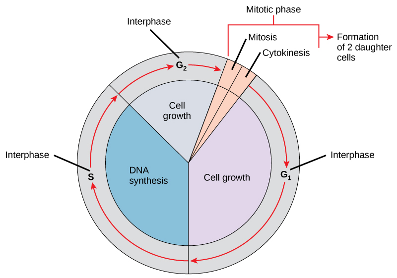
The cell cycle is an ordered series of events involving jail cell growth and cell division that produces two new daughter cells. Cells on the path to jail cell division go on through a serial of precisely timed and carefully regulated stages of growth, DNA replication, and nuclear and cytoplasmic partition that ultimately produces two identical (clone) cells. The cell wheel has 2 major phases: interphase and the mitotic phase ((Effigy)). During interphase, the cell grows and DNA is replicated. During the mitotic stage, the replicated Dna and cytoplasmic contents are separated, and the prison cell cytoplasm is typically partitioned by a third process of the cell cycle called cytokinesis. We should note, nevertheless, that interphase and mitosis (kayrokinesis) may take place without cytokinesis, in which case cells with multiple nuclei (multinucleate cells) are produced.
The jail cell cycle in multicellular organisms consists of interphase and the mitotic phase. During interphase, the cell grows and the nuclear Dna is duplicated. Interphase is followed by the mitotic phase. During the mitotic phase, the duplicated chromosomes are segregated and distributed into girl nuclei. Following mitosis, the cytoplasm is usually divided as well past cytokinesis, resulting in two genetically identical girl cells.
Interphase
During interphase, the cell undergoes normal growth processes while also preparing for cell division. In society for a cell to motility from interphase into the mitotic stage, many internal and external conditions must be met. The iii stages of interphase are called Yardane, S, and K2.
10001 Phase (First Gap)
The starting time stage of interphase is chosen the G1 phase (starting time gap) because, from a microscopic point of view, footling change is visible. However, during the Gi stage, the prison cell is quite active at the biochemical level. The cell is accumulating the building blocks of chromosomal Deoxyribonucleic acid and the associated proteins every bit well as accumulating sufficient free energy reserves to complete the job of replicating each chromosome in the nucleus.
S Phase (Synthesis of Deoxyribonucleic acid)
Throughout interphase, nuclear Dna remains in a semi-condensed chromatin configuration. In the South phase, DNA replication can go on through the mechanisms that result in the formation of identical pairs of DNA molecules—sister chromatids—that are firmly fastened to the centromeric region. The centrosome is as well duplicated during the S phase. The two centrosomes of homologous chromosomes will give rising to the mitotic spindle, the apparatus that orchestrates the movement of chromosomes during mitosis. For example, roughly at the center of each animal cell, the centrosomes are associated with a pair of rod-like objects, the centrioles, which are positioned at right angles to each other. Centrioles assistance organize cell partition. We should annotation, however, that centrioles are not present in the centrosomes of other eukaryotic organisms, such as plants and nearly fungi.
Thousand2 Phase (Second Gap)
In the G2 phase, the cell replenishes its energy stores and synthesizes proteins necessary for chromosome manipulation and movement. Some cell organelles are duplicated, and the cytoskeleton is dismantled to provide resources for the mitotic stage. In that location may exist additional cell growth during Gtwo. The concluding preparations for the mitotic phase must be completed before the cell is able to enter the first stage of mitosis.
The Mitotic Phase
The mitotic stage is a multistep process during which the duplicated chromosomes are aligned, separated, and motion into two new, identical daughter cells. The first portion of the mitotic phase is called karyokinesis, or nuclear division. As we have only seen, the second portion of the mitotic phase (and often viewed as a process carve up from and following mitosis) is called cytokinesis—the physical separation of the cytoplasmic components into the two daughter cells.
Link to Learning
Revisit the stages of mitosis at this site.
Karyokinesis (Mitosis)
Karyokinesis, too known as mitosis, is divided into a series of phases—prophase, prometaphase, metaphase, anaphase, and telophase—that consequence in the division of the prison cell nucleus ((Figure)).
Karyokinesis (or mitosis) is divided into 5 stages—prophase, prometaphase, metaphase, anaphase, and telophase. We should note that this is a continuous procedure, and that the divisions betwixt the stages are not detached. The pictures at the bottom were taken by fluorescence microscopy (hence, the black background) of cells artificially stained by fluorescent dyes: blue fluorescence indicates Deoxyribonucleic acid (chromosomes) and green fluorescence indicates microtubules (spindle apparatus). (credit "mitosis drawings": modification of work by Mariana Ruiz Villareal; credit "micrographs": modification of piece of work by Roy van Heesbeen; credit "cytokinesis micrograph": Wadsworth Eye/New York Country Section of Health; scale-bar information from Matt Russell)
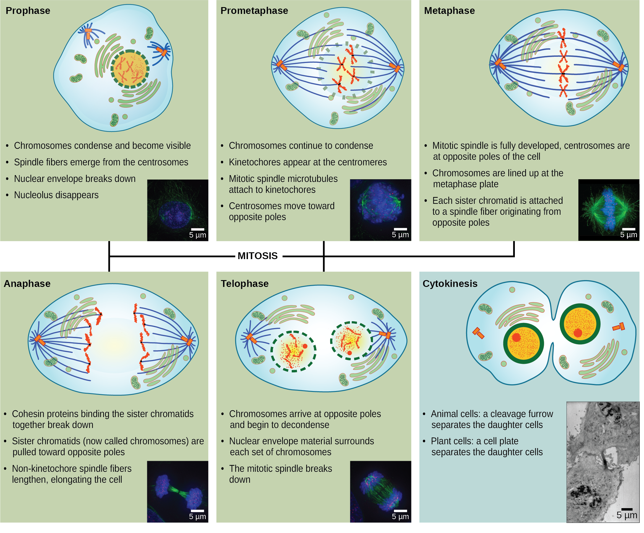
Prophase (the "first phase"): the nuclear envelope starts to dissociate into small vesicles, and the membranous organelles (such every bit the Golgi complex [Golgi apparatus] and the endoplasmic reticulum), fragment and disperse toward the periphery of the prison cell. The nucleolus disappears (disperses) too, and the centrosomes brainstorm to move to opposite poles of the jail cell. Microtubules that volition form the mitotic spindle extend between the centrosomes, pushing them farther autonomously as the microtubule fibers lengthen. The sister chromatids begin to coil more tightly with the aid of condensin proteins and now become visible under a lite microscope.
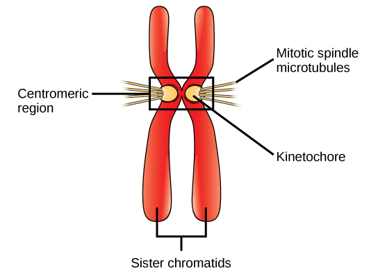
Prometaphase (the "first change phase"): Many processes that began in prophase continue to advance. The remnants of the nuclear envelope fragment further, and the mitotic spindle continues to develop equally more microtubules assemble and stretch across the length of the former nuclear expanse. Chromosomes go even more condensed and discrete. Each sister chromatid develops a protein structure called a kinetochore in its centromeric region ((Effigy)). The proteins of the kinetochore attract and bind to the mitotic spindle microtubules. As the spindle microtubules extend from the centrosomes, some of these microtubules come into contact with and firmly bind to the kinetochores. Once a mitotic fiber attaches to a chromosome, the chromosome will be oriented until the kinetochores of sister chromatids face the contrary poles. Eventually, all the sis chromatids volition be attached via their kinetochores to microtubules from opposing poles. Spindle microtubules that do not engage the chromosomes are called polar microtubules. These microtubules overlap each other midway between the two poles and contribute to cell elongation. Astral microtubules are located near the poles, assistance in spindle orientation, and are required for the regulation of mitosis.
During prometaphase, mitotic spindle microtubules from reverse poles adhere to each sister chromatid at the kinetochore. In anaphase, the connectedness between the sis chromatids breaks down, and the microtubules pull the chromosomes toward opposite poles.
Metaphase (the "change phase"): All the chromosomes are aligned in a airplane called the metaphase plate, or the equatorial plane, roughly midway betwixt the two poles of the cell. The sis chromatids are all the same tightly attached to each other by cohesin proteins. At this time, the chromosomes are maximally condensed.
Anaphase ("upward stage"): The cohesin proteins degrade, and the sis chromatids separate at the centromere. Each chromatid, at present called a single chromosome, is pulled rapidly toward the centrosome to which its microtubule is attached. The prison cell becomes visibly elongated (oval shaped) as the polar microtubules slide confronting each other at the metaphase plate where they overlap.
Telophase (the "distance phase"): the chromosomes attain the opposite poles and begin to decondense (unravel), relaxing over again into a stretched-out chromatin configuration. The mitotic spindles are depolymerized into tubulin monomers that will be used to assemble cytoskeletal components for each daughter cell. Nuclear envelopes form effectually the chromosomes, and nucleosomes appear within the nuclear expanse.
Cytokinesis
Cytokinesis, or "cell motion," is sometimes viewed every bit the 2nd main stage of the mitotic phase, during which cell division is completed via the physical separation of the cytoplasmic components into two daughter cells Withal, as we have seen earlier, cytokinesis can too be viewed as a separate phase, which may or may non take place following mitosis. If cytokinesis does take place, cell division is non complete until the cell components take been apportioned and completely separated into the 2 daughter cells. Although the stages of mitosis are similar for most eukaryotes, the process of cytokinesis is quite dissimilar for eukaryotes that accept cell walls, such as plant cells.
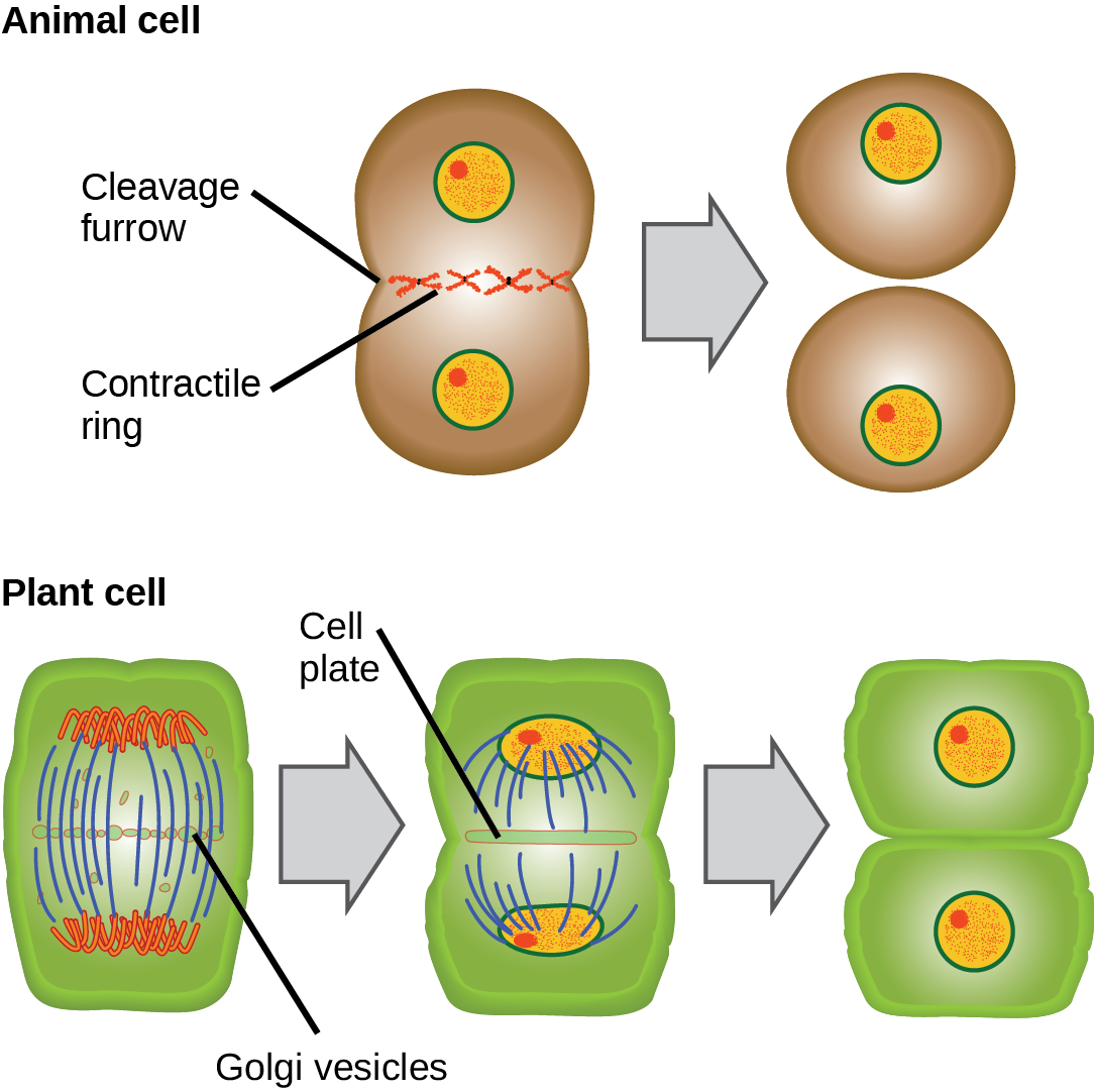
In animal cells, cytokinesis typically starts during late anaphase. A contractile ring composed of actin filaments forms just inside the plasma membrane at the one-time metaphase plate. The actin filaments pull the equator of the jail cell inward, forming a fissure. This fissure is called the cleavage furrow. The furrow deepens as the actin band contracts, and eventually the membrane is cleaved in two ((Effigy)).
In plant cells, a new cell wall must form betwixt the daughter cells. During interphase, the Golgi apparatus accumulates enzymes, structural proteins, and glucose molecules prior to breaking into vesicles and dispersing throughout the dividing cell. During telophase, these Golgi vesicles are transported on microtubules to form a phragmoplast (a vesicular structure) at the metaphase plate. There, the vesicles fuse and coalesce from the heart toward the cell walls; this structure is chosen a cell plate. As more vesicles fuse, the cell plate enlarges until information technology merges with the prison cell walls at the periphery of the cell. Enzymes utilise the glucose that has accumulated betwixt the membrane layers to build a new cell wall. The Golgi membranes become parts of the plasma membrane on either side of the new jail cell wall ((Effigy)).
During cytokinesis in animal cells, a ring of actin filaments forms at the metaphase plate. The ring contracts, forming a cleavage furrow, which divides the cell in 2. In plant cells, Golgi vesicles coalesce at the former metaphase plate, forming a phragmoplast. A cell plate formed by the fusion of the vesicles of the phragmoplast grows from the middle toward the jail cell walls, and the membranes of the vesicles fuse to form a plasma membrane that divides the cell in two.
G0 Phase
Not all cells adhere to the classic cell-cycle pattern in which a newly formed daughter cell immediately enters the preparatory phases of interphase, closely followed past the mitotic phase, and cytokinesis. Cells in G0 phase are not actively preparing to carve up. The cell is in a quiescent (inactive) phase that occurs when cells exit the prison cell cycle. Some cells enter Grand0 temporarily due to environmental conditions such as availability of nutrients, or stimulation by growth factors. The prison cell will remain in this phase until conditions improve or until an external betoken triggers the onset of G1. Other cells that never or rarely separate, such as mature cardiac muscle and nerve cells, remain in M0 permanently.
Scientific Method Connexion
Determine the Fourth dimension Spent in Cell-Bike Stages
Trouble: How long does a cell spend in interphase compared to each stage of mitosis?
Background: A prepared microscope slide of whitefish blastula cantankerous-sections will show cells arrested in various stages of the cell cycle. (Note: It is non visually possible to separate the stages of interphase from each other, but the mitotic stages are readily identifiable.) If 100 cells are examined, the number of cells in each identifiable cell-cycle stage will requite an estimate of the fourth dimension information technology takes for the jail cell to complete that stage.
Problem Statement: Given the events included in all of interphase and those that have place in each stage of mitosis, approximate the length of each stage based on a 24-hr cell cycle. Before proceeding, country your hypothesis.
Exam your hypothesis: Test your hypothesis by doing the following:
- Identify a fixed and stained microscope slide of whitefish blastula cross-sections nether the scanning objective of a lite microscope.
- Locate and focus on 1 of the sections using the low-power objective of your microscope. Detect that the department is a circle equanimous of dozens of closely packed individual cells.
- Switch to the medium-power objective and refocus. With this objective, individual cells are clearly visible, just the chromosomes will still be very pocket-sized.
-
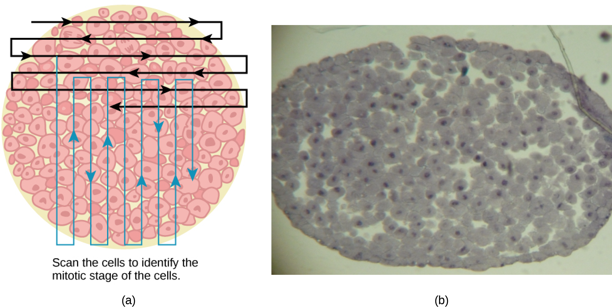
Switch to the loftier-power objective and slowly motility the slide left to right, and up and down to view all the cells in the section ((Effigy)). Equally you scan, you will find that most of the cells are non undergoing mitosis only are in the interphase menstruation of the prison cell cycle.
Slowly scan whitefish blastula cells with the high-power objective equally illustrated in image (a) to identify their mitotic stage. (b) A microscopic image of the scanned cells is shown. (credit "micrograph": modification of piece of work by Linda Flora; calibration-bar data from Matt Russell)
- Practice identifying the various stages of the cell wheel, using the drawings of the stages as a guide ((Figure)).
- Once you are confident about your identification, brainstorm to record the stage of each prison cell yous run into equally you scan left to right, and pinnacle to bottom across the blastula section.
- Keep a tally of your observations and end when you achieve 100 cells identified.
- The larger the sample size (total number of cells counted), the more accurate the results. If possible, assemble and record group data prior to calculating percentages and making estimates.
Record your observations: Brand a tabular array similar to (Figure) within which to tape your observations.
| Results of Cell Stage Identification | |||
|---|---|---|---|
| Stage or Stage | Individual Totals | Group Totals | Percent |
| Interphase | |||
| Prophase | |||
| Metaphase | |||
| Anaphase | |||
| Telophase | |||
| Cytokinesis | |||
| Totals | 100 | 100 | 100 percent |
Clarify your data/report your results: To find the length of time whitefish blastula cells spend in each stage, multiply the percent (recorded as a decimal) by 24 hours. Make a table similar to (Figure) to illustrate your information.
| Judge of Cell Phase Length | ||
|---|---|---|
| Phase or Stage | Percent | Time in Hours |
| Interphase | ||
| Prophase | ||
| Metaphase | ||
| Anaphase | ||
| Telophase | ||
| Cytokinesis | ||
Draw a conclusion: Did your results support your estimated times? Were any of the outcomes unexpected? If so, hash out those events in that phase that may have contributed to the calculated fourth dimension.
Section Summary
The cell wheel is an orderly sequence of events. Cells on the path to cell division go on through a series of precisely timed and carefully regulated stages. In eukaryotes, the cell wheel consists of a long preparatory period, called interphase, during which chromosomes are replicated. Interphase is divided into Grand1, S, and G2 phases. The mitotic phase begins with karyokinesis (mitosis), which consists of five stages: prophase, prometaphase, metaphase, anaphase, and telophase. The final stage of the cell division process, and sometimes viewed equally the final stage of the mitotic phase, is cytokinesis, during which the cytoplasmic components of the daughter cells are separated either by an actin ring (animate being cells) or past cell plate formation (plant cells).
Review Questions
Chromosomes are duplicated during what stage of the prison cell wheel?
- Thoui phase
- South phase
- prophase
- prometaphase
B
Which of the following events does not occur during some stages of interphase?
- DNA duplication
- organelle duplication
- increase in cell size
- separation of sis chromatids
D
The mitotic spindles arise from which cell construction?
- centromere
- centrosome
- kinetochore
- cleavage furrow
B
Zipper of the mitotic spindle fibers to the kinetochores is a characteristic of which stage of mitosis?
- prophase
- prometaphase
- metaphase
- anaphase
B
Unpacking of chromosomes and the germination of a new nuclear envelope is a feature of which stage of mitosis?
- prometaphase
- metaphase
- anaphase
- telophase
D
Separation of the sister chromatids is a characteristic of which stage of mitosis?
- prometaphase
- metaphase
- anaphase
- telophase
C
The chromosomes go visible under a light microscope during which stage of mitosis?
- prophase
- prometaphase
- metaphase
- anaphase
A
The fusing of Golgi vesicles at the metaphase plate of dividing establish cells forms what construction?
- prison cell plate
- actin band
- cleavage furrow
- mitotic spindle
A
(Figure) Which of the following is the correct order of events in mitosis?
- Sis chromatids line up at the metaphase plate. The kinetochore becomes attached to the mitotic spindle. The nucleus reforms and the jail cell divides. Cohesin proteins break downward and the sister chromatids divide.
- The kinetochore becomes attached to the mitotic spindle. Cohesin proteins suspension down and the sis chromatids separate. Sis chromatids line up at the metaphase plate. The nucleus reforms and the cell divides.
- The kinetochore becomes attached to the cohesin proteins. Sis chromatids line upwardly at the metaphase plate. The kinetochore breaks downward and the sister chromatids separate. The nucleus reforms and the cell divides.
- The kinetochore becomes attached to the mitotic spindle. Sis chromatids line upwardly at the metaphase plate. Cohesin proteins break downward and the sister chromatids separate. The nucleus reforms and the jail cell divides.
(Figure) D. The kinetochore becomes attached to the mitotic spindle. Sister chromatids line up at the metaphase plate. Cohesin proteins pause downwardly and the sister chromatids divide. The nucleus reforms and the cell divides.
Critical Thinking Questions
Briefly draw the events that occur in each phase of interphase.
During Gi, the cell increases in size, the genomic DNA is assessed for damage, and the cell stockpiles energy reserves and the components to synthesize Dna. During the Due south stage, the chromosomes, the centrosomes, and the centrioles (animal cells) duplicate. During the Chiliadtwo phase, the cell recovers from the S phase, continues to grow, duplicates some organelles, and dismantles other organelles.
Chemotherapy drugs such as vincristine (derived from Madagascar periwinkle plants) and colchicine (derived from autumn crocus plants) disrupt mitosis by binding to tubulin (the subunit of microtubules) and interfering with microtubule assembly and disassembly. Exactly what mitotic construction is targeted past these drugs and what effect would that have on cell division?
The mitotic spindle is formed of microtubules. Microtubules are polymers of the protein tubulin; therefore, it is the mitotic spindle that is disrupted by these drugs. Without a functional mitotic spindle, the chromosomes volition not exist sorted or separated during mitosis. The cell will arrest in mitosis and dice.
Describe the similarities and differences between the cytokinesis mechanisms constitute in animal cells versus those in found cells.
At that place are very few similarities between animal cell and establish cell cytokinesis. In animal cells, a ring of actin fibers is formed around the periphery of the cell at the former metaphase plate (cleavage furrow). The actin ring contracts inwards, pulling the plasma membrane toward the middle of the cell until the cell is pinched in two. In plant cells, a new prison cell wall must be formed between the daughter cells. Due to the rigid cell walls of the parent cell, wrinkle of the heart of the cell is not possible. Instead, a phragmoplast outset forms. Subsequently, a cell plate is formed in the middle of the cell at the erstwhile metaphase plate. The cell plate is formed from Golgi vesicles that contain enzymes, proteins, and glucose. The vesicles fuse and the enzymes build a new cell wall from the proteins and glucose. The jail cell plate grows toward and eventually fuses with the cell wall of the parent cell.
List some reasons why a cell that has merely completed cytokinesis might enter the Thou0 phase instead of the Thousand1 phase.
Many cells temporarily enter G0 until they reach maturity. Some cells are but triggered to enter Gi when the organism needs to increase that item cell type. Some cells only reproduce post-obit an injury to the tissue. Some cells never split one time they reach maturity.
What cell-bicycle events will be affected in a cell that produces mutated (not-functional) cohesin protein?
If cohesin is not functional, chromosomes are not packaged later DNA replication in the S stage of interphase. Information technology is likely that the proteins of the centromeric region, such as the kinetochore, would not grade. Fifty-fifty if the mitotic spindle fibers could attach to the chromatids without packing, the chromosomes would not be sorted or separated during mitosis.
Glossary
- anaphase
- stage of mitosis during which sister chromatids are separated from each other
- cell bicycle
- ordered series of events involving jail cell growth and cell division that produces two new daughter cells
- cell plate
- structure formed during plant cell cytokinesis by Golgi vesicles, forming a temporary construction (phragmoplast) and fusing at the metaphase plate; ultimately leads to the formation of cell walls that separate the two girl cells
- centriole
- rod-similar construction constructed of microtubules at the heart of each beast cell centrosome
- cleavage furrow
- constriction formed past an actin band during cytokinesis in fauna cells that leads to cytoplasmic partition
- condensin
- proteins that help sister chromatids coil during prophase
- cytokinesis
- division of the cytoplasm following mitosis that forms 2 girl cells.
- Thousand0 phase
- distinct from the 10001 phase of interphase; a cell in G0 is not preparing to divide
- Gone stage
- (likewise, commencement gap) first phase of interphase centered on jail cell growth during mitosis
- G2 phase
- (besides, 2nd gap) 3rd phase of interphase during which the jail cell undergoes final preparations for mitosis
- interphase
- period of the cell cycle leading upwardly to mitosis; includes G1, S, and 1000two phases (the acting period between two consecutive cell divisions)
- karyokinesis
- mitotic nuclear division
- kinetochore
- poly peptide construction associated with the centromere of each sis chromatid that attracts and binds spindle microtubules during prometaphase
- metaphase plate
- equatorial airplane midway between the two poles of a jail cell where the chromosomes marshal during metaphase
- metaphase
- phase of mitosis during which chromosomes are aligned at the metaphase plate
- mitosis
- (as well, karyokinesis) menses of the cell wheel during which the duplicated chromosomes are separated into identical nuclei; includes prophase, prometaphase, metaphase, anaphase, and telophase
- mitotic phase
- period of the cell wheel during which duplicated chromosomes are distributed into two nuclei and cytoplasmic contents are divided; includes karyokinesis (mitosis) and cytokinesis
- mitotic spindle
- apparatus composed of microtubules that orchestrates the movement of chromosomes during mitosis
- prometaphase
- stage of mitosis during which the nuclear membrane breaks down and mitotic spindle fibers attach to kinetochores
- prophase
- stage of mitosis during which chromosomes condense and the mitotic spindle begins to form
- quiescent
- refers to a cell that is performing normal prison cell functions and has not initiated preparations for cell division
- S phase
- second, or synthesis, phase of interphase during which Deoxyribonucleic acid replication occurs
- telophase
- phase of mitosis during which chromosomes go far at contrary poles, decondense, and are surrounded by a new nuclear envelope
Source: https://opentextbc.ca/biology2eopenstax/chapter/the-cell-cycle/
Posted by: mcleodsallithere.blogspot.com
0 Response to "How Do Animal Cells Complete The Cell Cycle"
Post a Comment